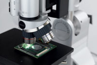
Microscopy has made some enormous changes in the last several years. The microscopy industry has adapted by developing new features and improving the ergonomics of their products. Source: Aven Inc.
If the microscope cannot “talk” to the computer or camera or software, it may find itself out of a job. All components of the system must be connected for best results.
As Tom Calahan, product manager at Carl Zeiss MicroImaging Inc. (Thornwood, NY) says, microscopy today is all about integrated solutions. In the past, there would be a microscope, possibly with a computer attached, and a camera or imaging device, but that did not mean the instruments all worked together. “All the equipment wasn’t well integrated,” Calahan says. “What the customer is looking for nowadays-and the industry is moving toward-is complete integrated solutions with the camera, software and microscope all talking in unison.”
When taking an image of the sample, some systems now have the ability to not only store the image, but also to know the conditions in place when the image was taken, along with the magnification, resolution and optical technique used. All of that information goes back to the computer, providing the operators with more control over the system. By archiving that information, the operator is able to stay in touch with remote colleagues or use the same imaging technique two months later. This has practical real-world applications in today’s manufacturing environment.
“One quality control group in California wants to do the same thing they are doing in New York,” Calahan says, and this system allows them to replicate the conditions. “All this is tied into the integration of the digital imaging system with the microscope. It is no longer separate components not talking to each other. Integration makes everything run smoother and better in the long run,” he says.

Manufacturers strive to make new technology available and easy to use. Source: Aven Inc.
The Evolution
The evolution of microscopy and digital imaging began years ago, but within the past three to four years, integrated solutions really started to come together. Several factors led to this change, particularly camera developments and capabilities of the microscope.With cameras, operators are moving toward digital. They can get higher-resolution, with a greater dynamic range, and comparable quality to film. In addition, the speed on the computer side means that operators can transfer files in 30 seconds to a group on the other side of the world, Calahan says.
But cameras do not deserve all the credit: enhancements in microscopes also led to this change. Microscopes today are becoming more motorized and encoded. Imaging software controls the camera and microscope, allowing the system to take a picture of the image and later repeat the same technique.
In Calahan’s almost 32 years at Zeiss, the technology has taken off. “I think over the years there was a slower start, but in the mid-90s, digital imaging really started to accelerate,” he says. “Technology was one of the drivers-it made digital imaging what it is today and what it will be in the future, and brought along with it the microscopy side of things, with the control and motorization.
“Definitely, the quality of imaging has grown over the years,” says Calahan, “and it will continue in the future. The end customer solution is what everybody is striving for.”
Eventually this may lead to improved motorization and larger imaging devices, depending on customer demand. “The customer is in the driving seat at this point,” Calahan says.
Trends
Mike Shahpurwala, marketing director at Aven Inc. (Ann Arbor, MI), says digital imaging is the biggest trend in microscopy these days.“Everything is going toward digital imaging,” Shahpurwala says. The new technology allows operators to take that image, share it and view it on a computer or LCD monitor.
Before the advent of digital imaging, operators attached a Polaroid camera to the top of the microscope and took a picture. With the CCD or CMOS cameras, they can now view it on a screen.
The technology produces high-quality images, and the megapixels keep increasing, Shahpurwala says. Operators now are able to bring that image into a computer and do a detailed analysis of the image, and then save or archive it.
The field of microscopy is a constant work in progress, as companies adapt to fit the marketplace and develop new features. One such adaptation is the third port on the microscope.
These third ports have been around for a while, but now can be used with digital cameras. This allows for documentation, and lets the operator e-mail the image. Some microscope ports can be used with any camera on it. Others are standardized, and work with one camera type.
“They’re finding a way to keep their market space. There’s room for everybody,” says Greg Hollows, vision integration partners coordinator at Edmund Optics (Barrington, NJ). “At least for now. There’s always some disruptive technology.”
Technology keeps moving forward, Hollows says. “Ten years ago getting an image into a computer was a three-day process,” Hollows says. Today, this can be done by simply plugging a cable into the computer.
Technological advances are not without bumps in the road. Cheaper is not always better when purchasing these products, Hollows warns. The goal should be to simply have the desired results at the end of the day.
In the future, Hollows predicts, products will be faster with higher resolution, and the digital camera market will continue to get better, though it will eventually cap out somewhere. Hollows also emphasizes that demand will drive development.

Product Development
Though companies may have the ability to create many different features, they would be wise to listen to what customers want.“Microscopes are not sold as one-size-fits-all,” Calahan says. “We talk to the customer, ask what they’re doing and what’s needed down the road.”
Customers run the gamut, and the company aims to provide a specific solution for each application.
Based on customer demand, Aven has developed a system incorporating all the different components into a single piece of equipment. Previously, in order to get the image the operator had to attach a CCD camera or a digital camera to the trinocular port, take the image from the camera to a PC or monitor, and have the PC or monitor nearby. By incorporating all of that into one system, the operator does not need a separate computer.
“We have seen customers wanting a product of this nature,” Shahpurwala says, and Aven expects to release the product in the first quarter of next year.
Portable microscopy is another trend on its way, says Shahpurwala. Operators will be able to carry the microscope, and still be able to take a very high magnification image. This capability is particularly useful when examining metal parts in a large jet engine where operators previously could not take a microscope. Thus, moving a unit from one station to another becomes just a matter of unplugging it.
Microscopy will start seeing a lot of integrated systems, and new products aim to fit this need.
“Customers want systems that are simpler to use,” Shahpurwala says. “They don’t want to ask, ‘Will this camera work? This software? Do I need an adapter?’ By incorporating it all into one system, they don’t have to worry about it.”
These requests illustrate the ever-changing world of microscopy. Though technology may evolve, the industry has not stood still. Hollows credits the microscopy industry for adapting well to customers’ needs and improving the features and ergonomics of their products. “Microscopy is still a big area,” Hollows says. “They are always upping the bar there and they’ve adapted well to a variety of industries.”Q
Videoscope Basics
Increasingly videoscopes are being used for imaging applications. They display magnified images on monitors or flat panel displays.A typical videoscope consists of a video lens coupled to a camera connected to a PC or monitor mounted to a stand. A lighting system, either LED or fiber optic, also may be added to enhance image quality.
A variety of video lenses are available in macro and micro formats for a range of magnification requirements. Because of the large selection and configurations available with video lenses, a large range of field of views, magnification ranges and working distances are available.
Videoscopes provide a more ergonomic solution for imaging applications and can improve operator comfort and productivity.
br> Choosing the correct video lens for an application will require the following consideration:
- Field of View: The area of the object that will be viewed on the monitor
- Resolution: The smallest possible resolvable feature of the object
- Depth of Field Requirement: Maximum object depth needed for focus
- Minimum and Maximum working distance
When choosing between a videoscope and a microscope with camera there are a few key points to remember:
4A microscope system provides the operator with the ability to see a true 3-D image when looking through the microscope eyepieces. This may be very important for some applications. The same 3-D depth of field is not possible on a videoscope because the image is being projected on a 2-D screen or monitor.
4Videoscopes tend to have a higher magnification range and more lens options. Most stereomicroscopes go up to 200X magnification, while a videoscope is able to go 1,400X and higher.
4A videoscope reduces eye fatigue for the operator.
The ideal system would be an easy-to-use microscope and videoscope merged into a plug-and-play unit.
After selecting the lens system and camera, the next step is to choose the correct stand and lighting system for the application.
- - Mike Shahpurwala, Aven Inc.
Optical Microscopes
If planning to use an optical microscope for image acquisition, the following is needed:- An optical scope with an additional port loaded on the body for mounting a camera, often called a trinocular microscope. Note: most CCD or CMOS cameras have either C or CS mounts. Most manufacturers provide adapters for mounting these cameras or their ports may be threaded to accommodate a camera.
- Choose the correct camera for the application. A wide range is available from analog to digital. Because microscopic images have an intrinsic limiting resolution, avoid using a noisy, high-resolution detector for image acquisition. A more modest detector with larger pixels often can produce higher quality images because of reduced noise. In addition, a lower resolution detector often will have a significantly higher acquisition rate permitting the observation of faster events. Conversely, if the object is motionless, one may want to acquire the image at highest possible resolution.
- Choose the right software for the application. There are a variety of software packages available from a multitude of vendors as either stand-alone packages or supplied with cameras. Typical analysis includes edge detection, counting similar objects, measurement of an object, creating image masks, annotation and report generation.
- - Mike Shahpurwala, Aven Inc.
Tech tips
For more information, about the companies mentioned in this article, visit:
- Aven Inc., www.aveninc.com
Carl Zeiss MicroImaging, www.zeiss.com/micro
Edmund Optics, www.edmundoptics.com

