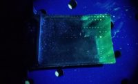
A feasibility study looks into micro-radiographic identification and eddy current detection.
We conducted a feasibility study for the detection of micron sized (greater than or equal to 50 micron) inclusions in rolled mild steel material of a nominal thickness of 0.035 inch. The purpose of this study was to determine if a real time nondestructive testing method could be applied that would lend itself to in-situ testing conditions that exist in a hot rolling mill. After a preliminary evaluation it appeared that an eddy current technique might be used in providing real-time, in process data during the production of rolled sheet material that would provide useful metrics for monitoring product process control as evidenced by the distribution and relative quantity and size of naturally occurring trapped inclusions resulting from a hot mill process. The attributes of eddy current testing that were attractive included the need to acquire data under rapid surface scanning requirements and provide volumetric test results lending to high speed data analysis.
In approaching this task, it was determined that two test elements would be needed to support the process of the study. The first challenge was to nondestructively find, identify and locate anomalous indications that might be contained in sample material as would be representative of a less dense than base material inclusion. The second challenge was to demonstrate a surface scan eddy current technique that could reasonably match with reproducible results the detection of the radiographically identified inclusions.
In order to meet the first challenge the radiographic nondestructive volumetric testing method was selected in the manner of micro-radiography. Although micro-radiography is not considered a typical routine industrial test method, it has been increasingly used in recent years for nondestructive testing of small electronic components (both 2-D and 3-D) for the defense and aerospace industry and for 3-D evaluation of specialty castings and unique composite and metallic components. Its application in the medical community is well known. There are several OEMs of micro-radiographic systems available today and in use for industrial applications.
The first phase for the evaluation of samples (nominal size of 12 inches by 14 inches by 0.035 inches) was to use the micro-radiography technique with a sensitivity and resolution capable of identifying the x-y location of micron sized indications in the base metal matrix. Material samples were selected at random from a single “run” of sheet material and a visual surface inspection was performed to identify surface indications. The six samples were radiographed using an X-ray inspection system with an effective X-ray focal spot of 0.0000394 inch. The 2-D digital imaging method used an image intensifier with a pixel resolution of 4,000 pixels per square inch.
In typical radiographic context, the image unsharpness and detail sensitivity was more than sufficient to resolve indications of 50 microns or greater. The radiographic test results presented images wherein the measured sizes of identified inclusions ranged from about 40 microns to 120 microns in profile. The X-ray image produced progressive and indexed images, each image of a 0.1 inch by 0.1 inch area. Each sample received a 100% scan. Images were produced with and without a superimposed, computer generated grid. The resultant images were captured in JPEG format for the purpose of image interpretations and analysis. (Typical radiographic images with a representative indication are shown above.) One must keep in mind that the image processing with this X-ray inspection system produces an image with reverse density contrast, meaning lighter appearing inclusions are representative of less dense regions than the base material.
The images were evaluated for all six samples, representing in excess of 20,000 individual images, and a single representative sample (referenced herein as the “master” sample) that exhibited typical and multiple inclusion type indications was selected for further testing. All of the identified indications within the single sample were recorded with the X-Y coordinate as given by the image identification index. In order to provide a zero reference location on each plate, the plates were marked with an indent on a corner to represent the X0-Y0 location. Each image was digitally recorded by the computerized system and indexed from the zero reference marks in increments of 0.1 inch.
The second phase of the evaluation was to determine the feasibility of detection of the indications found in the first phase using eddy current testing coupled with a saturated magnetic field. Eddy current testing was performed using a system along with eddy current probes, with data capture and analysis performed with the windows based software via PC LAN connection to the system for delivery of real time data display, data management and storage. This system was selected due to its advanced computer based software system and extensive signal evaluation options. Consideration was made in selecting a eddy current system that was ideally suited to operate on a production line with remote Windows PC or central operator panel as an integral part of a process control system as might be required for on-line, in-situ testing in a rolling mill with a software end user application.
In order to perform the eddy current testing on the samples (sections of actually rolled material) a fixture was fabricated that would compress the rolled (slight curvature), samples and provide a flat surface for surface scanning. The fixture included an eddy current probe holder, probe proximity and adjustable permanent magnets mounts, and x-y scanning guide frame that allowed for X-Y position manual scans of the plate sample.
Repeated eddy current testing (scans) was performed on the “master sample” for a known one inch width by 12 inches length section of the sample for which there were nine radiographically recorded individual indications ranging in size from about 40 to 100 microns in nominal diameter. This region of the master sample was selected since the adjacent area on either side of the one-inch strip was relatively free of other noted radiographic noted indications. Multiple scans were performed at various instrumentation settings until a reasonable and clear set of return eddy current signals were recorded. The resulting eddy current critical test parameters were for the frequency set for 30 kilohertz with an instrumentation gain of 43 to 50 decibels. The total scan length for eddy current test results were matched to the radiographic identified indications for the nine internal inclusions contained in the one inch wide and 11 inches length.
It is possible to view the results of the image vs. comparative eddy current data. The single image frame represents one identified inclusion within the 0.1 inch by 0.1 inch area of the sample. The scans represent two separate scans of a one-inch wide strip containing multiple indications, as represented by the single identified inclusion. A correlation was made based on the actual location of the indication in the sample, and the single “peak” recorded as typical for that indication. There were a total of nine similar inclusions contained in the one-inch wide strip of the about 12 inches of scan length.
Confirming scans were made of the calibration test sample to confirm that the eddy current test parameters would produce a signal response from the three flat bottom drilled holes when the test sample was tested from the opposing side of the sample plate containing the flat bottom indications.
Extensive testing was performed on the “master sample” and whereas the data was consistent, not all of the radiographically recorded indications were detected by the eddy current testing. Based on the number of repeated scans performed in both the X and Y direction, one can state that approximately 80% of the “known” locations for internal indications produced a concise and noted eddy current response signal.
In summary, it appears feasible that an eddy current testing using a saturated magnetic field allows detection of the micron sized internal indications (inclusions) in the material and per the test parameters briefly described herein. An extensive amount of testing was performed and reproducible results obtained for the eddy current recognition of internal inclusions. It should be noted that with increased scanning speed the eddy current signal response appeared to become more distinct, which is expected. Whereas the test did even approximate the 600 feet per minutes (fpm) of actual longitudinal speed as would be seen in production the compact system is in use successfully in other applications in a wire drawing operation where the speed has been measured at up to 18,000 fpm. Further testing is recommended and should be followed by physical metallographic examination to determine the type and nature of the indications identified by the micro radiographic method and characterized by the eddy current testing. In addition, further test and data comparisons need to be conducted in continuing confirmation of this feasibility testing.


