Triboluminescence: Enabling Major Advances in XRF
Triboluminescent X-ray generation has had a profound impact on XRF technologies.
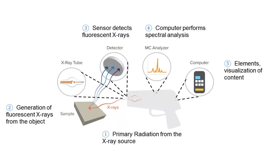
Figure 1. Schematic of XRF Process.
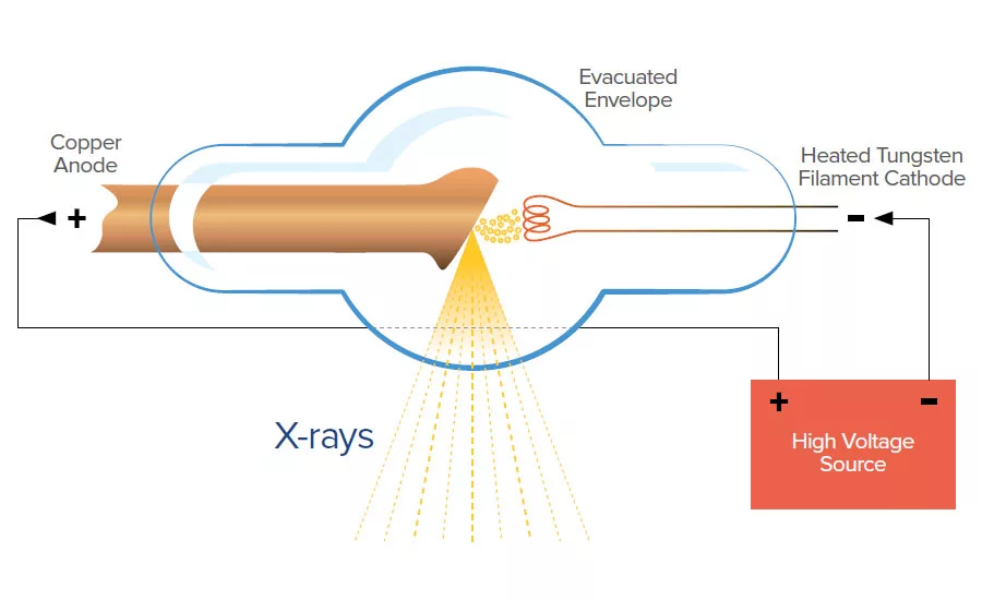
Figure 2. Conventional X-ray Tube.
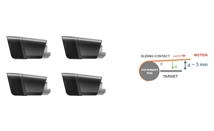
Figure 3. Field-replaceable X-ray tubes enabled by novel triboluminescent X-ray technology.
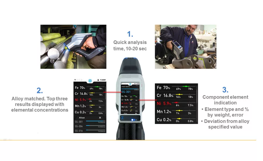
Figure 4. Spectrum Data Indication
The first choice for use in an increasing range of applications, such as medicine, mineralogy, agriculture, manufacturing, construction, geology, and archeology, X-ray fluorescence (XRF) spectrometry and analysis has undergone significant advancement over the past century. XRF spectrometry is a nondestructive, non-intrusive analytical technique that can be used to determine the chemical composition of metals, glass, ceramics, and other materials in various physical states such as solids, powders, coatings, and liquids. Many samples can be analyzed with little to no preparation, making XRF an easy quality check compared to other analysis techniques. Cost and size minimizations are among the most important advancements expanding the use of XRF instruments worldwide. Two decades ago the first handheld XRF (HHXRF) technology became commercially available, enabling a vital transition from stationary benchtop XRF instrumentation to mobile devices enabling spectrometry and analysis in the field.
However, despite the numerous advancements, the heart of all XRF technology—the generation of X-rays—has been stagnant since 1895. At the end of the 19th century, Wilhelm Conrad Roentgen was studying cathode rays emitted from a high-voltage gaseous discharge tube. He happened to notice that a barium-platinocyanide screen encased in a cardboard box that was lying near the experiment would fluoresce whenever the tube was in operation, despite being surrounded by opaque material. Thus, X-rays were discovered! Then, in 1909, Charles Glover Barkla discovered a connection between X-rays radiating from a sample and the atomic weight of the sample, establishing the foundations of XRF technology. However, nearly 40 years passed before the first XRF spectrometer was built by Herbert Friedman and Laverne Stanfield Birks, Jr., paving the way for commercial use of XRF technology. Figure 1 depicts the spectrometry and analysis process for XRF, a nondestructive measurement.
Materials emit photons as a result of being bombarded with enough high-energy X-rays. That is X-ray fluorescence. The types of atoms in the material affect the energy they radiate back. Each atom in the periodic table emits a different, unique energy. These discrete energy values are what an XRF spectrometer relies on to identify what the sample is composed of and in what ratios.
Fragile, bulky, and expensive high-voltage gaseous discharge tubes remain essential to X-ray technologies, harking back to 1895. A high voltage source creates a potential across the vacuum tube. A filament, typically tungsten, is heated and emits electrons that accelerate towards the positively charged anode. Upon collision, the electrons’ energy is converted into X-rays that are directed out of the vacuum tube towards the sample.
The detectors and the computers around XRF have all undergone dramatic changes, but the core technology has remained unchanged since its discovery. That is, until the discovery of novel triboluminescent X-ray technology by a team of physicists at UCLA in 2008. Backed by DARPA funding, the physicists were expanding upon an obscure observation reported by Russian researchers in the 1980s: crushing certain types of rocks in a vacuum produces luminescence in the X-ray energy range. So, what is triboluminescence? It’s the phenomenon of creating light through mechanical action such as pulling apart, ripping, scratching, crushing, or rubbing different materials. Known by ancient civilizations, this phenomenon can be observed when crushing sugar crystals and peeling adhesive tapes, but only recently has the potential for generating high-voltage discharge been harnessed.
Triboluminescent X-ray generation has a profound impact on XRF technologies, enabling cost and size reductions at a scale not possible with conventional technology. It is now possible to rely on the triboelectric effect caused by mechanically pulling materials together and apart to discharge enough electrons at the target anode. An X-ray beam sufficient for successful XRF spectrometry is generated, obviating the need for a fragile glass tube; a bulky, dangerous high-voltage power supply; and the supporting components found in conventional XRF devices. An electric motor, battery, switch, microcontroller, and a low-voltage connector replace the need for inverters, transformers, and high-voltage control systems. The remaining sub-systems of HHXRF spectrometers remain the same. With the high-voltage power supply eliminated, it is now safe for the user to change the X-ray tube without needing extensive safety training and certification. No longer does a company have to experience downtime because their HHXRF spectrometer is in the shop for costly routine tube replacement. Field replacement of the X-ray source is now a reality, for zero downtime.
Triboluminescent X-ray generators are the primary innovation driving down the cost of entry for HHXRF spectrometers by almost half, and creating many new uses for HHXRF. In addition to novel developments in the generation of X-rays, dual detector technology for HHXRF is now available. Real time calibration is realized, with no need for manual user calibration.
The first users that will benefit from this advancement are machining, manufacturing, and scrap recycling operators. With the reduction of prohibitive entry level pricing, many more users will find use out of HHXRF, such as the aerospace, automotive, military, and medical industries. In fact, companies within those sectors are now requiring verification of materials beyond the certificate. Triboluminescent X-ray technology enables users to not only trust their certificates, but verify them, opening more doors for business and reducing risk across the board. Anyone with concern over the quality of their materials, from incoming shipments to internal processing to the final product, should consider low-cost triboluminescent HHXRF technology.
Looking for a reprint of this article?
From high-res PDFs to custom plaques, order your copy today!


