Consider the magnification, resolution, contrast and measurement when buying a microscope.

FocalSpot releases the Optilia Flexia BGA/SMT video inspection microscope. Source: FocalSpot Inc.


Several factors-advanced materials research, device miniaturization and nanotechnology-have contributed to the growing need for greater precision and versatility in microscopic imaging.

When selecting a microscope, consider the magnification, resolution, contrast, depth of field, field of view, illumination method, degree of automation and type of image produced. The three basic types: electron, confocal and compound microscopes, each offer distinct advantages for the operator.

Also, new products are continually introduced, such as the confocal laser scanning microscope, the scanning tunneling microscope, the atomic force microscope, and the video microscope. Thus, no matter what the application, there is no shortage of microscopes and imaging solutions available.


The DG-3 portable digital microscope can be used in the field or factory. Source: Aven Inc.
FocalSpot Inc. (www.focalspot.com) offers the Optilia Flexia BGA/SMT video inspection microscope. The Flexia is a versatile electrostatic discharge (ESD)-protected video microscope for look-down or look-under visual inspection of solder joints on assembled printed circuit board's (PCB's) components, solder/flux mixtures, bare circuit cards/pads, cables/connectors, wire bonds and stencils. Flexia can be set up in seconds with either a look-down macro zoom lens (1 to 60X) or look-under (4X zoom) side viewing ball grid array (BGA) lens. It can be connected directly to a handheld monitor, PC or laptop via a high speed USB 2.0 frame grabber. Imaging applications include digital image capture, video recording and optical measurements. The system is optimized for lead-free inspection and provides robust data management and documentation tools. The side-viewing lens of the Flexia offers the capability to look-under and inspect BGAs, µBGAs, CSP and flip chip packages with minimal package stand off. The Flexia BGA lens captures sharp images of the soldered balls under the BGA package up to 12 millimeters deep and down to ~0.05 millimeter stand off. The Flexia BGA lens allows operators to perform failure analysis-opens, bridging, micro-cracking, surface defects-and confirm solder ball shape, reflow compression, wetting and flux conditions, and other visual characteristics of SMT/BGA components. Optional Picasara software provides facilities to measure bump height, radius, wetting angles, while Picasara's database and reporting environment make possible the development of case examples for inspection verification reference, training materials and other documentation requirements.


The LSM 5 DUO laser scanning microscope combines two microscopes into a single instrument. Source: Carl Zeiss
Aven Inc. (www.aveninc.com) is the U.S. distributor for Scalar Scopes, the manufacturer of the DG-3 portable digital microscope. Surfaces are magnified with its LED illuminated lenses-from 25X to 1000X-and then imaged by its 2.3 million pixel CCD sensor. NASA, Boeing, the United Space Alliance, Florida Power & Light, and some major automakers are currently using DG units. An engineer with Boeing at Kennedy Space Center reported that they are now using the digital microscope for inspection of bolt holes, banding screens, corrosion and braided flex hoses. Boeing has also purchased the 25 to 200X par focal zoom lens. This lens can be used as a touch-and-view lens or it can provide about a 1/2-inch working distance when its touch collar is removed. The zoom lens allows operators to scan an area at 25X, bulls eye a defect or area of interest, and then zoom to 200X without moving the lens. The strength of the DG-3 is its portability-it can be used in the field or factory, in a fuselage, on animals, pipes, PCBs and nearly any material requiring quality control or nondestructive testing. The product weighs 500 grams and works off a lithium-Ion battery. With its touch-and-view technology, operators can capture images on site, which helps minimize downtime and maximizes profits. Images are recorded on an industry standard CF card.


The STM6 (left) and SZ61 (right) from Olympus Micro-Imaging offer flexibility in illumination and imaging. Source: Olympus Micro-Imaging
Carl Zeiss (www.zeiss.com) offers versatility in laser scanning microscopes with the LSM 5 DUO that combines two scanning modules in a single confocal microscope. It combines two confocal laser scanning microscopes, the LSM 510 META and LSM 5 LIVE, to form a single instrument. The fast line-scanning module of the LSM 5 LIVE and the precise point-scanning module of the LSM 510 META are integrated in the new workstation.

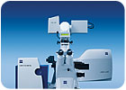
The two independent scanner groups provide flexibility in optical sample micromanipulation for FLIP or FRAP applications or when utilizing photoactivateable and photoconvertible variants of fluorescent proteins. Dynamic processes up to the kilohertz range occur using the system's fast acquisition capabilities. Meanwhile, multiple META channels make time-saving, specimen-preserving spectral imaging possible. The LSM 5 DUO is well-suited to a wide range of live cell imaging applications, allowing researchers to probe deeper into the cell's molecular processes. Developmental studies can be carried out at high speed and in 3-D resolution, even with low-magnification lenses. Physiological measurements can be perfectly matched to biological timescales or spectral properties. The META detector enables fluorochromes with high overlapping emission spectra to be precisely separated using its emission fingerprinting technology. Photomanipulation experiments involving pixel-precise laser irradiation, such as FRAP, FLIP, FLAP, photo-activation or conversion and uncaging, can be conducted at high-resolution timescales. Fast 4-D image acquisition enables dynamic structures to be monitored simultaneously.


The new JSM-6390/6490 series comprises five models. Source: Jeol USA
Olympus Micro-Imaging's SZ series of zoom stereomicroscopes are available in two microscope designs. The Greenough design offers a lightweight and compact microscope while providing 3-D viewing and an outstanding depth of field. Galilean design models have highly corrected optical systems and attain higher magnifications, while offering modularity and a large range of illumination choices. All models, with the proper adapters, can accept a variety of digital cameras for image archiving. The new STM series of measuring microscopes includes models for fast inspection of large samples and the high-precision STM6 offers the most compact body in this class. These scopes feature motorized focusing, advanced UIS optics, and a high-rigidity body with integrated counters.


Magnetic roller feet allow the microscope to fit snuggly around the item to be inspected. Source: Pyser-SGI
Jeol USA (www.jeolusa.com), a supplier of scientific and analytical instruments, introduces a series of high-resolution tungsten scanning electron microscopes (SEMs) with multiple live image displays, streamlined graphical interface and improved low kilovolt operation. The new SEM series enables simultaneous observation of up to three different images-secondary electron, backscattered electron, and digital camera-on-screen measurement, and smart settings for simplified functionality. Secondary electron resolution is 3 nanometers at 30 kilovolts, 8 nanometers at 3 kilovolts, and 15 nanometers at 1 kilovolt, and magnification ranges from 5 to 300,000X.

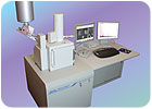
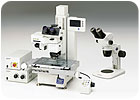
New electron optics enhance both general purpose imaging as well as analysis at the nanoscale. The new JSM-6390/6490 series comprises five models, offering a choice of low-vacuum operation, three stage sizes for specimens up to 12 inches in size, and goniometer stage axis control. In addition, JEOL USA offers a range of specially designed sample holders for its U.S. customers. JEOL offers a line of electron microscopes, including field emission SEMs, transmission electron microscopes (TEMs), and several new instruments designed for sample preparation, including an argon beam cross section polisher and a new single-column focused ion beam system.

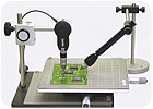
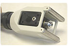
Surface inspection of circular objects can be problematic because of the difficulty of keeping the optical microscope stable.


Magnetic roller feet allow the microscope to fit snuggly around the item to be inspected. Source: Pyser-SGI

Pyser-SGI (www.pyser-sgi.com) has overcome this problem with the introduction of the portable tube scope. Magnetic roller feet allow the microscope to fit snuggly around the item to be inspected. It can then be easily focused on the area of interest and the through lens illumination, powered by a battery pack, gives bright clear images even on the darkest surfaces. Magnifications from 100 to 400X can be supplied and a calibrated graticule is fitted in the eyepiece to provide direct measurements. The scopes robust metal construction and quality glass optics ensure that it will give long-term performance. The polished wooden box houses the unit when not in use.