Materials Analysis
Do Ions Matter in Cut Face Quality?
Learn more about the impact of ion type on FIB cut face quality.
.webp?t=1763650467&width=1080)
.webp?t=1763650467&width=1080)
High-quality FIB-cut faces are essential for accurate analysis in materials research, supporting reliable imaging, defect detection, and cross-sectional studies. FIB works like a nanoscale “sandblaster,” directing a concentrated ion beam onto a material’s surface to sputter away atoms and reveal a new face. This enables researchers to cut into a sample with precision, expose hidden internal structures, and prepare site-specific regions for imaging and analysis at the micro- and nanoscale.
What defines a high-quality cut face?
The goal of any FIB workflow is to create a cross section that reveals the material as it is, without introducing artifacts or mechanical/chemical distortions. If the cross section fails to represent the material accurately, it could have unwanted effects on the analysis process.
One major pain point in FIB milling is curtaining, which appears as wavy or grooved patterns on the surface of the milled cross-section, resembling the folds of a curtain. It often occurs when a sample has layers of different materials of varying densities, causing the ion beams to remove some areas faster than others. Curtaining can massively impact the quality of analysis as it distorts the real structure of the material and can obscure fine features such as thin layers, grain boundaries or defects.
The top boundary of the cross section must be clean, straight and well-defined. For accurate measurement of features like thin films, interfaces or microstructures, well-defined edges are essential — especially when comparing layers in multilayered materials. Round or smeared edges can make it difficult to determine exact dimensions, leading to errors in interpretation or calculations.
Therefore, a high-quality cut face should meet several criteria: it must be smooth and curtain free with no streak-like milling artifacts, it should have a sharp and well-defined top edge with a clear boundary at the surface and should have a damage-free surface layer that isn’t compromised by extra defects introduced by the ion beam.
While these fundamentals define what quality looks like in practice, the specific choice of ion plays a decisive role in whether these standards can be achieved.
Examining ion types
The ability to mill and serial-section material using FIB milling inherently depends on the interactions between the ion beam and the solid sample. Milling occurs through physical sputtering, where incoming ions knock atoms away from the surface, but the outcome is shaped by several variables. The bombarding ion species, its energy, the crystallographic orientation of the target and the angle of incidence of the ion beams, all critically influence whether the resulting cut face is smooth, artifact-free and representative of the true structure of the material.
Gallium has long served as a favored choice for FIB systems, as it provides excellent beam stability and precise control. It is extracted from a liquid metal ion source (LMIS), which provides excellent beam stability and control. However, gallium can chemically interact with certain materials, leaving behind contamination, and its relatively low maximum beam current restricts throughput when working with larger samples or volumes.
By contrast, other ion sources such as xenon, nitrogen, argon and oxygen are generated using inductively coupled plasma (ICP) technology — collectively known as Plasma-FIB (PFIB) systems. PFIB instruments can deliver much higher ion currents than LMIS, making them particularly efficient for high-throughput milling while still offering flexibility for precision cross-sectioning.
Xenon, for example, offers an alternative that addresses some of these limitations. With a higher atomic mass, xenon produces a much greater sputter yield than gallium, enabling it to remove material at a faster rate. This makes it particularly effective for coarse milling or when working on larger sample areas. Beyond speed, xenon is also regarded as one of the best for preparing transmission electron microscopy (TEM) lamellas, particularly when minimizing structural damage is a priority.
Argon is another option, given it is inert and relatively light. These properties allow it to minimize surface damage and avoid ion implantation. As a result, argon is well suited for preparing delicate, ultra-thin samples where maintaining the integrity of the atomic structure is essential. On the other hand, nitrogen plays a much more specialized role in FIB applications. In addition to its milling capabilities, nitrogen ions can chemically interact with target materials to form nitrides on the surface. This process can increase hardness, making it attractive when surface modification is as important as quality cross sectioning.
Oxygen, on the other hand, is particularly effective for soft, non-conductive materials such as polymers and tissues, which are difficult to mill cleanly at high rates. Oxygen PFIB produces smooth, curtain-free surfaces even in challenging samples. Where gallium and xenon can risk producing curtaining artifacts in these materials, oxygen ions create more uniform cuts that reveal fine structural details without heavy post-processing — making them the best choice for composites research, failure analysis and life sciences applications.
So, does ion choice matter?
Together, these examples underline a central point: ion choice is not incidental, but decisive. Selecting the right ion type for the material and application at hand is essential to achieving the high-quality cross sections on which accurate imaging and reliable analysis depend.
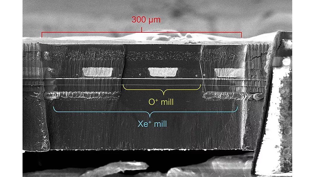
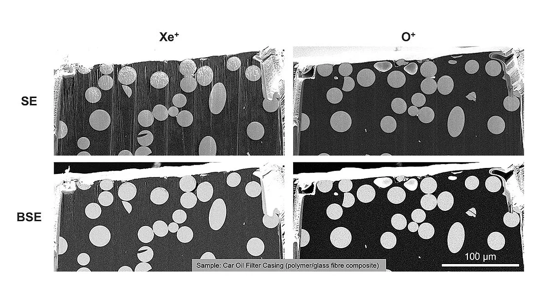
One illustrative case, carried out using Thermo Fisher Scientific equipment, comes from the study of a glass-fiber reinforced nylon 66 composite, a material widely used in automotive components such as oil filter casings. The composite contains about one-third glass fibers, each roughly 15 microns in diameter, embedded in a nylon matrix. This structure gives the material its stiffness and resistance to temperature extremes, chemicals and moisture, but also makes it challenging to prepare clean cross sections for analysis.
To examine the composite, researchers first used micro computed tomography (microCT) to locate regions of interest containing fibers and sparsely distributed defects. Guided by the reconstructed 3D data, they then employed a correlative workflow to target these regions for serial sectioning with focused ion beam milling. Both xenon and oxygen ions were tested under identical conditions. The comparison was striking: xenon milling produced noticeable curtaining, while oxygen PFIB created smooth, curtain-free surfaces that revealed fine microstructural details down to the nanometer scale.
The outcome underscored the importance of ion choice. While xenon remained usable at lower beam currents, oxygen plasma proved far more effective for this polymer-based composite, delivering cleaner sections and more reliable data. This example highlights how tailoring ion type to the material at hand can directly improve both cut-face quality and the efficiency of the overall analysis.
Looking for a reprint of this article?
From high-res PDFs to custom plaques, order your copy today!


