Optimize Synthetic Aperture Focusing
Ultrasonic testing of materials can be done in several ways using different types of instruments, ranging from thickness gages for localized measurements to multi-axis automated C-Scan scanners for imaging of complex parts. While these instruments can satisfy the needs of most applications, some situations require advanced signal analysis techniques in order to maximize the information collected from the tested structure.
One of the problems related to structure inspection is the correct identification of the ultrasonic echoes and time-of-flight related to defects. Differentiating between echoes from defects and from the nondefected surrounding areas of the material can be challenging, particularly when complex geometries are involved. In addition, time-of-flight or phase information of a single echo produced by a defect is usually insufficient to correctly determine the origin of this echo in space, and completing defect sizing is impossible. However, multiple echoes acquired from different probe locations along with numeric or synthetic focusing could contain enough information to localize defects in materials and display clearer cross-sectional B-scans and full C-Scan images.
Basic Principles
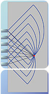
A single ultrasonic echo can be represented by multiple properties such as its center frequency, bandwidth, amplitude or energy, as well as time-of-flight or phase. All of these characteristics can provide valuable information about defect size or material attenuation, for example. However, when it comes to defect localization, only time-of-flight or phase are considered valuable information. Unfortunately, ultrasonic beams are never perfectly unidirectional and a defect can be the source of an echo for multiple probe positions. A single A-Scan is therefore generally insufficient to determine the exact location of a defect because a given time-of-flight can only provide information about the absolute distance from the transducer. However, a correct comparison of the echoes of a defect recorded from multiple probe positions can provide this information.The principles of synthetic aperture focusing rely on the same basis as radar or phased-array imaging. In the case of the radar, sweeps of electromagnetic waves are sent in the space to cover a predetermined aperture. The electromagnetic waves are then reflected if they meet an electrically leading surface or if an obstacle is in the propagation direction. The position of this obstacle is then obtained by comparing the echoes recorded within the aperture.
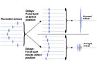
In synthetic aperture focusing of ultrasounds, a transducer emitting ultrasonic waves is moved along the test piece in pulse-echo mode to create a scanning aperture. If a defect is present in the aperture, echoes can be generated for multiple scanning positions, allowing to locate the defect. While a perfect transducer may be unidirectional to easily locate defects, synthetic apertures are optimized by beam divergence. Defect location is obtained by correctly delaying the echoes recorded from multiple probe locations in order to synchronize their phase and to average the resulting A-Scans. The area of interest of the structure under evaluation is separated into a matrix of positions and the delays are calculated in order to focus at each individual pixel of the B-Scan or C-Scan.
If a defect is present within a reconstructed pixel, the phase of the corresponding echoes match and a constructive summation, or constructive interference, occurs. On the other hand, if the focal spot is located in a nondefected pixel, the echoes are not in phase and the summation will be randomly constructive and destructive. The result of this process is a pixelized representation of the structure, where focusing is done on each pixel and defects are identified by high amplitude of echoes. Synthetic focusing on B-Scans-time vs. probe position-results in a depth vs. probe position image, while a complete X-Y-Z positioning is done with synthetic focusing on C-Scans.
The principles on which this focusing method is based are compliant with small defect detection. In fact, the ideal defect for an optimal image reconstruction is a small one. The whole phase matching process of multiple detections requires the defect to reflect ultrasonic energy toward the transducer. Small defects respond in such a way, mainly because of the wave diffraction phenomena.
However, large reflecting surfaces need to be either oriented correctly so that the waves reflect back in the correct direction, or rough enough to create diffuse reflections, for which orientation is not as important. For example, defects such as flaws will in most cases produce diffuse reflections and tip diffraction, both effects being favorable to synthetic focusing. On the other hand, surfaces which mainly produce specular reflections-reflection angle corresponding to the incidence angle-need to be properly oriented to reflect ultrasonic waves to the transducer, reducing the number of echoes that can be recorded for each point of this type of surface and the subsequent phase construction possibilities. This particular property has a certain interest since reflections from small defects can be discriminated from larger reflectors such as the boundaries of a tested piece.
Practical Synthetic Aperture Focusing
One of the best qualities of synthetic focusing is that it can be complementary to conventional ultrasonic scanning systems. As an analysis tool, synthetic aperture focusing adds focusing advantages to scanners not equipped with phased-array capabilities. For example, a conventional immersion scanner with a single element could be used to image a sample with the advantages of synthetic focusing. Furthermore, focusing at large depths using a single transducer can hardly be achieved and the use of common focused transducers is limited to certain depths. Synthetic aperture focusing allows high-resolution 2-D and 3-D imaging of structures using a single probe, which is suitable for situations where C-Scan imaging is involved.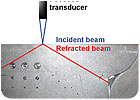
In general, synthetic aperture focusing also can be applied to multiple ultrasonic inspection fields. It is already widely used as a complement analysis tool for time-of-flight diffraction inspection. Hidden or inaccessible sections of structures also can be inspected and imaged when inspected using divergent transducers. Angle-beam inspection of welds also could benefit from synthetic focusing by considering the echoes originating from multiple wave paths within the structure to do focusing.
Advanced Testing
Advanced testing for aerospace components such as inspection of composite materials also could benefit from the use of focusing techniques. With a good knowledge of the wave velocity behavior within the composite structure-slowness curves-focusing is possible and smaller defects could be detected due to the better signal-to-noise ratio.In the past few years, the feasibility to use synthetic aperture focusing in the field of long range ultrasonic inspection-guided waves inspection-has been done successfully. This technique presents a good potential for the imaging of structures such as aircraft wing skins and hidden sections of fuselage components.
As radar technology collects reflected echoes and adds them in a manner to signal the presence of moving targets in the space, synthetic focusing collects echoes from targets within the inspection area and produces better image reconstruction of defects compared to conventional B-Scan or C-Scan images. Such clear images allow better target and defect interpretation and good signal-to-noise ratio. Applying the presented synthetic focusing technique to advanced inspection of material also could lead to higher lateral resolution in the inspected material, providing inspectors with better sizing of defects apparent in the produced synthetic B-Scan or C-Scan images. NDT
Looking for a reprint of this article?
From high-res PDFs to custom plaques, order your copy today!


